News

Research updates from MicroCD Labs.

Notes on microfluidics, diagnostics, product sourcing, and analysis tools that support research-use assay development.

Latest update MicroCD SonoMap research concept

Product research · July 25, 2026

Research concept — in development

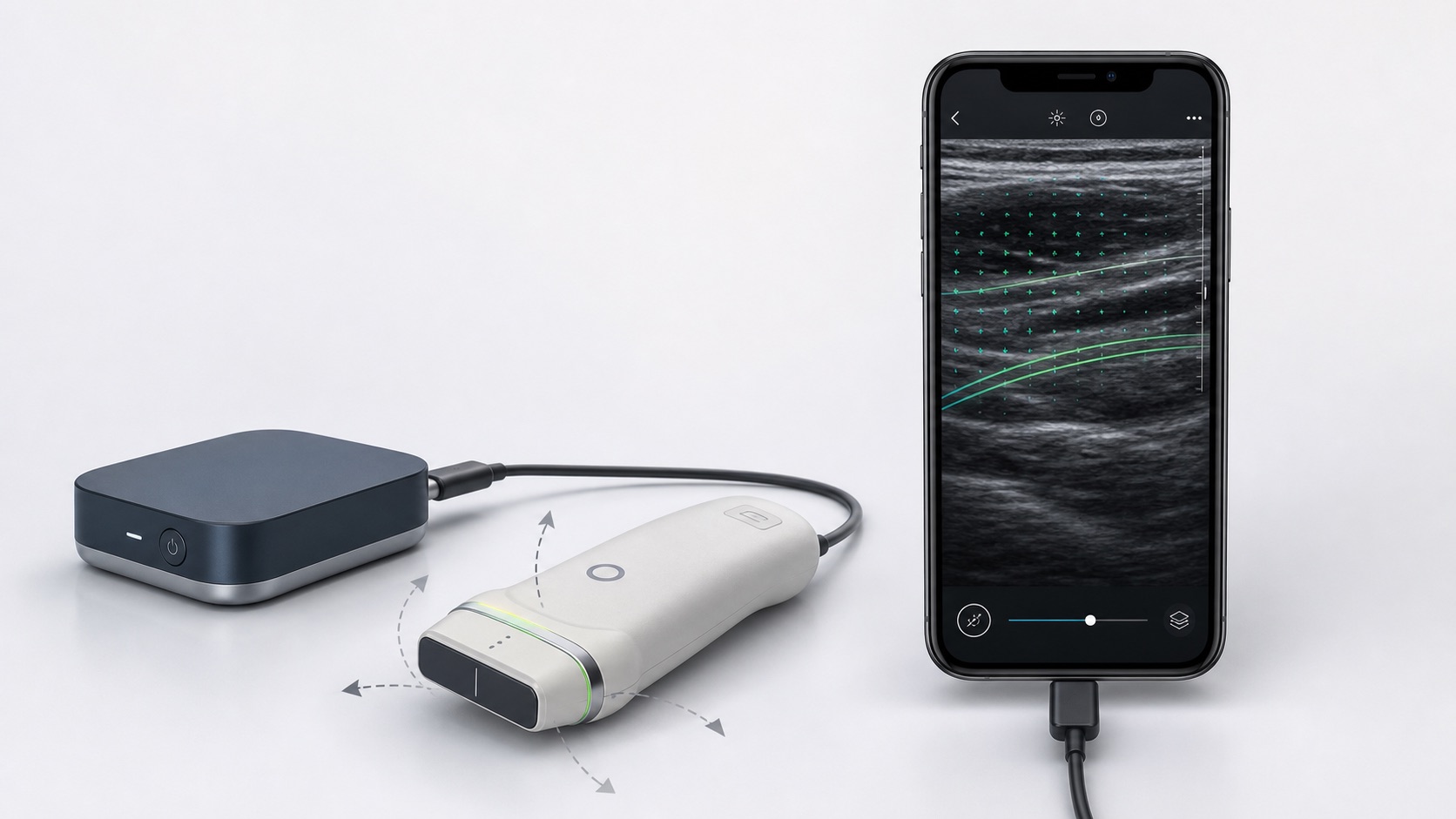
MicroCD SonoMap explores directional tissue mapping with compact ultrasound.

MicroCD Labs is defining an early-stage, phone-connected research platform for controlled multi-angle ultrasound acquisition, probe sensing, motion-aware reconstruction, and orientation-sensitive tissue maps.

Initial work is framed around bench feasibility, aligned-fibre phantoms, and non-clinical research data. SonoMap is not a finished or validated device and is not intended for diagnosis, treatment, patient monitoring, or clinical decision-making.

Concept rendering of a generic phone-connected ultrasound probe and external acquisition module displaying directional research vectors
Exploratory hardware and interface rendering. The design and imaging output are conceptual.

Field note · July 24, 2026

Organ-on-chip and 3D culture systems expand human-relevant testing.

Conventional two-dimensional cell cultures cannot reproduce every feature of living tissue. Three-dimensional culture and organ-on-chip systems provide controlled environments in which cells can organize, interact, and respond to flow or mechanical cues in ways that may better reflect aspects of human physiology.

Microfluidic organ-chip devices can support disease modeling, drug-response studies, and toxicity research using living human cells. These approaches may reduce reliance on some animal studies, but broader validation, standardization, and fit-for-purpose evidence are still needed before they can replace established models across drug development.

Selected references: Three-Dimensional Cell Culture Systems and Their Applications in Drug Discovery and Cell-Based Biosensors; Halfway between 2D and Animal Models: Are 3D Cultures the Ideal Tool to Study Cancer-Microenvironment Interactions?; and the Wyss Institute Human Organs-on-Chips program.

Why it matters

Microfluidic engineering is becoming part of the preclinical research toolkit.

Chip geometry, fluid control, materials, imaging, and repeatable operation all influence the quality of an organ-on-chip workflow.

Field note · July 24, 2026

Microfluidics is moving selected tests closer to patients.

By integrating sample preparation, fluid handling, reagents, and detection into compact devices, microfluidic systems can reduce the sample volume and equipment required for some diagnostic workflows. This is especially relevant where centralized laboratory infrastructure or trained operators are difficult to access.

University of Washington research led by Paul Yager has explored low-cost, instrument-minimized point-of-care tests using paper and porous-media fluidics. Wearable researchers, including teams associated with the University of Cincinnati, have also investigated microfluidic sweat collection as a less invasive route for measuring selected biomarkers.

These technologies remain application-specific. A sweat measurement is not automatically equivalent to a blood measurement, and smartphone imaging or control does not by itself establish analytical or clinical validity. Sampling, calibration, interference, stability, and intended-use validation remain central development questions.

Selected references: Enabling a Microfluidic Immunoassay for the Developing World by Integration of On-Card Dry Reagent Storage; Bioanalytical Devices: Technological Leap for Sweat Sensing; and A Wearable Patch for Continuous Analysis of Thermoregulatory Sweat at Rest.

Engineering focus

Useful devices must simplify the complete sample-to-result workflow.

Low-volume collection, dry-reagent storage, capillary flow, contamination control, readable outputs, and operator usability need to work as one system.

Platform launch · July 16, 2026

MicroCD LabOps launches for engineering documentation and traceability.

MicroCD Labs has launched MicroCD LabOps, a hosted workspace for scientific-hardware and diagnostic-development teams to organize engineering reports, supplier qualification, components, received lots, incoming inspections, and controlled review records.

The deployed platform now uses a connected Supabase database with organization-scoped access controls. LabOps is entering private beta: customer use remains subject to project-specific security, legal, validation, retention, and data-governance review, and the platform is not represented as a validated QMS, LIMS, electronic-signature, or clinical system.

MicroCD LabOps public beta homepage showing engineering documentation and traceability capabilities
MicroCD LabOps connects engineering reports, supplier records, components, lots, and inspections in one organization-scoped workspace.

Research update

Preprint available: kinetic image analysis for low-concentration assay detection.

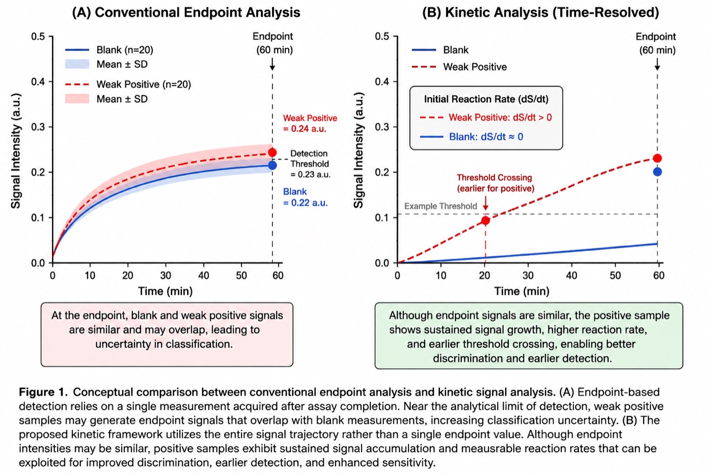
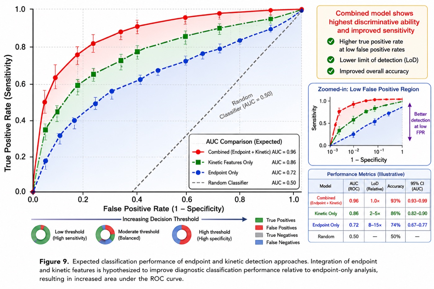
The latest MicroCD Labs-aligned manuscript, Improving Detection Limits in Capillary Diagnostic Assays Through Kinetic Image Analysis and Time-Resolved Signal Extraction, presents a framework for moving beyond single endpoint images in lateral-flow, dot-based, paper microfluidic, and lab-on-disc assays.

The work highlights time-series image acquisition, region-of-interest tracking, background correction, kinetic feature extraction, concentration estimation, and endpoint-versus-rate comparison for low-concentration diagnostic development.

Conceptual comparison of endpoint analysis and kinetic time-resolved analysis for weak positive capillary diagnostic assay signals
Endpoint signals can overlap near the detection limit; kinetic trajectories can reveal sustained signal growth earlier.
Workflow diagram for kinetic image acquisition, preprocessing, ROI detection, signal extraction, feature extraction, concentration prediction, and diagnostic decision
Time-resolved image analysis workflow
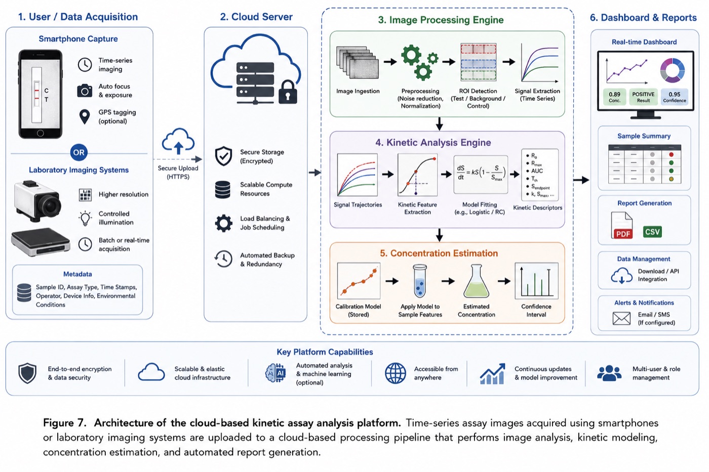
Cloud-based kinetic assay analysis platform architecture for smartphone capture, image processing, kinetic modeling, dashboard, and reports
Cloud analysis and reporting concept
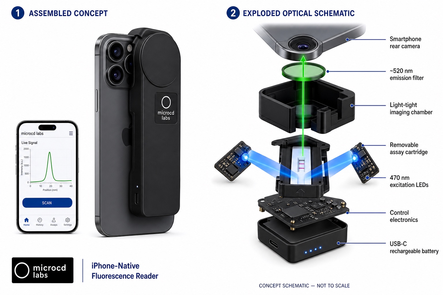
Concept schematic showing a smartphone fluorescence reader correctly aligned to a rear camera, with its emission filter, imaging chamber, assay cartridge, excitation LEDs, electronics, and battery
Exploratory reader architecture connecting smartphone image acquisition with time-resolved assay analysis. Concept schematic, not to scale; not a finished or validated device.