Directional tissue mapping
Estimate dominant orientation and regional coherence from multi-angle image sequences.

MicroCD SonoMap™
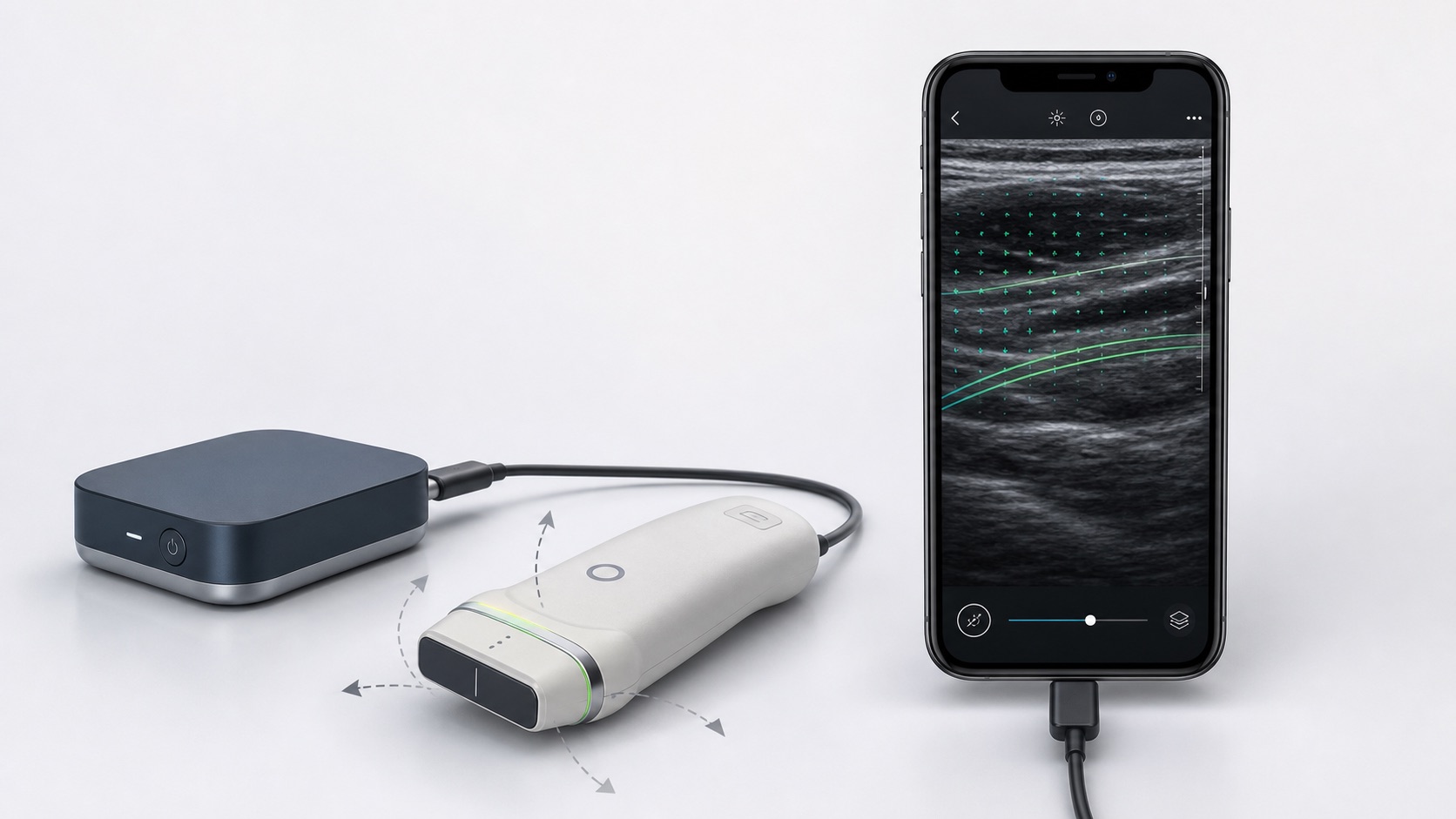
Research concept — in developmentA compact, phone-connected ultrasound research platform concept exploring how multi-angle acquisition, probe sensing, and computational reconstruction could reveal directional tissue architecture.
Research concept only. Not for diagnosis, treatment, patient monitoring, or clinical decision-making. No regulatory clearance, clinical validation, or performance specification has been established.
Research question
Conventional B-mode ultrasound is useful for visualizing structure, but tissue orientation can change with probe angle and may require manual interpretation. SonoMap is being framed around controlled multi-angle acquisition, position and force sensing, and reproducible computational analysis.
The intended output is a research map of orientation-sensitive image features, not MRI-equivalent tractography and not a diagnostic conclusion.
Proposed workflow
Each stage is designed to preserve acquisition context so researchers can inspect how the result was produced.
Capture conventional ultrasound frames or raw channel data across defined probe angles.
Record probe orientation, motion, and applied force to support repeatable acquisition.
Register the sequence and estimate orientation-sensitive texture, backscatter, or flow features.
Display maps, confidence indicators, acquisition metadata, and research-ready exports.
Potential research modes
Modes will be prioritized by bench evidence, data quality, and partner need rather than presented as finished capabilities.
Estimate dominant orientation and regional coherence from multi-angle image sequences.
Explore repeatable measures of aligned structures in phantoms and ex vivo samples.
Quantify angle-dependent changes in backscatter or related image features.
Investigate advanced flow workflows where suitable acquisition hardware and research protocols are available.
Use tracked movement and image registration to reduce inconsistencies across a sweep.
Preserve image series, sensor data, parameters, maps, and confidence measures for downstream analysis.
System architecture
The phone is envisioned as the control and review interface; ultrasound acquisition remains in dedicated external electronics.
Development plan
The first milestone is a reproducible research workflow, not a clinical product.
Research applications
Research basis
Published work has demonstrated automated estimation of muscle-fibre orientation in ultrasound images and has shown that transducer orientation affects fascicle measurements. Zhou & Zheng, 2008; Bolsterlee et al., 2016.
Backscatter tensor imaging research has also examined fibre direction through angle-dependent ultrasonic speckle coherence in anisotropic tissue. Papadacci et al., 2014.
Ultrasound localization microscopy is an emerging research method for microvascular visualization, but it has distinct acquisition requirements and remains separate from SonoMap's initial direction-mapping scope. Dencks & Schmitz, 2023.
These references inform the research direction. They do not validate MicroCD SonoMap or establish its performance.
Why MicroCD Labs
Instrument and workflow designTranslate an imaging hypothesis into a practical acquisition sequence, interface, and experimental plan.
Mechanical integrationCoordinate probe fixtures, sensor integration, electronics packaging, and prototype fabrication.
Data and verification planningDefine traceable datasets, comparison methods, acceptance criteria, and documented failure modes.
Research collaboration
We welcome conversations with ultrasound researchers, tissue-engineering teams, signal-processing specialists, transducer and electronics developers, and organizations evaluating research imaging workflows.
Please keep this initial message non-confidential. Do not include patient information, protected health information, unpublished data, or proprietary design details.